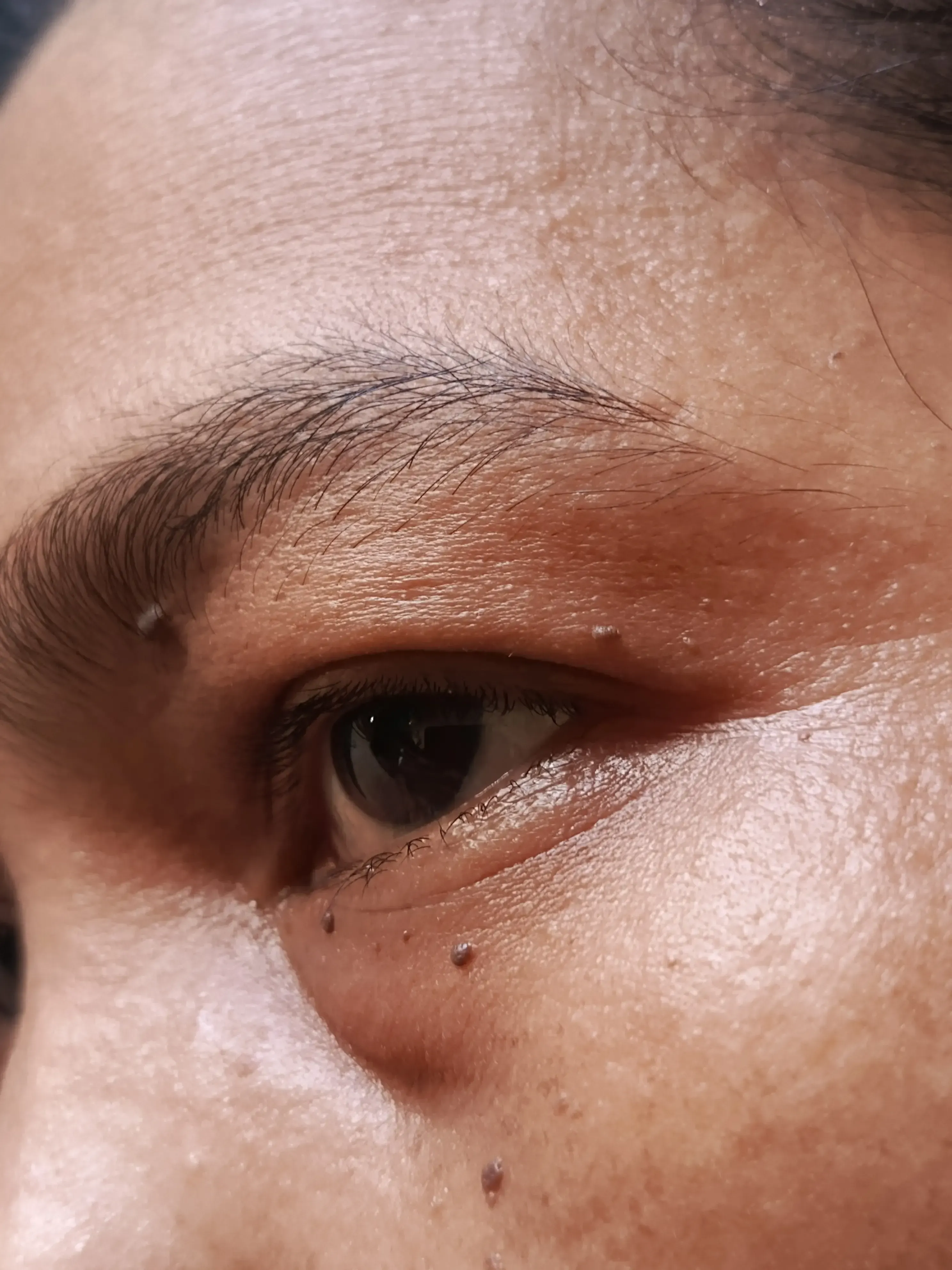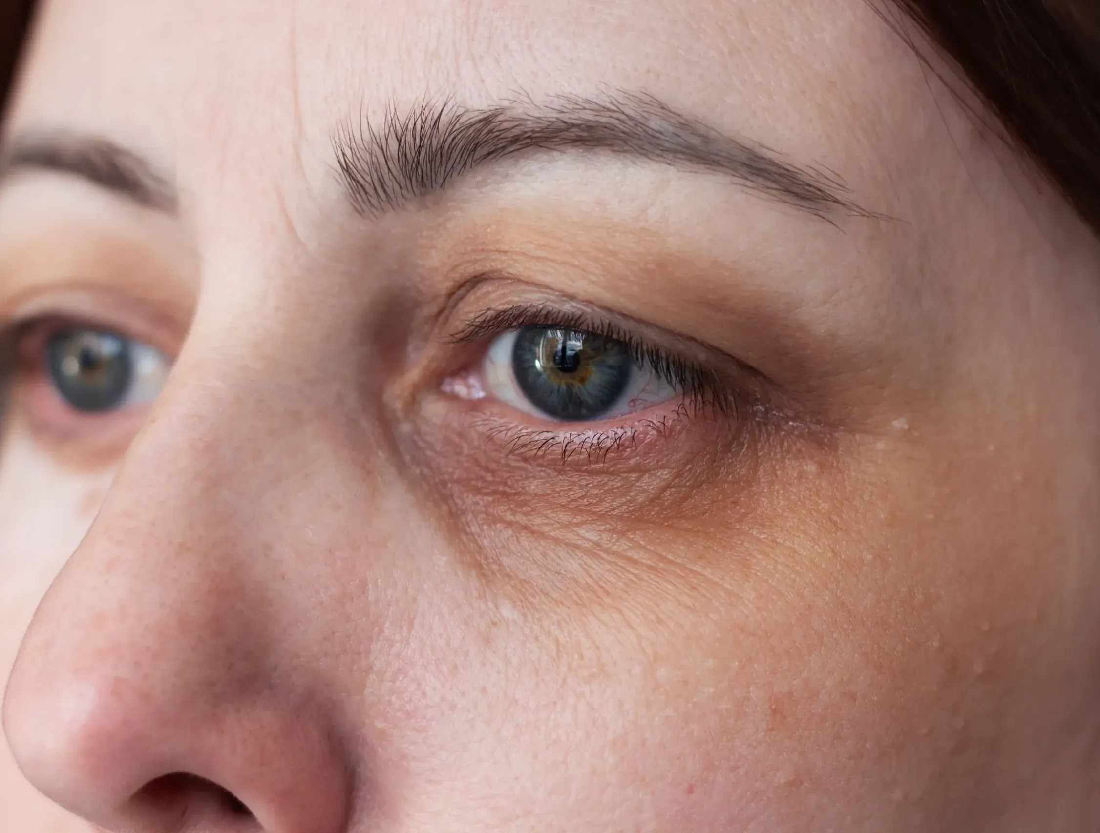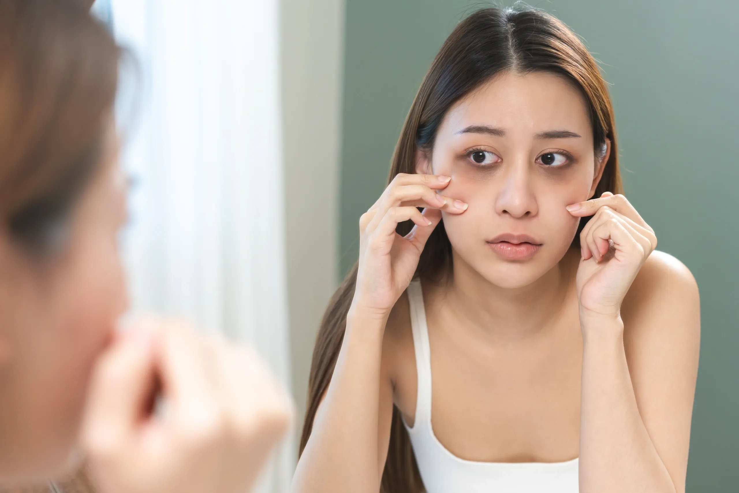

The skin beneath the lower eyelid is among the thinnest on the entire body, averaging just 0.5 mm thick compared with 2 mm on the cheeks. This anatomical reality means that blood vessels, melanin deposits, and underlying shadows are far more visible here than anywhere else on the face. The American Academy of Dermatology recognizes multiple contributing mechanisms, reflecting how this is a multifactorial condition rather than a single-cause problem.
Melanin production in the periorbital zone can increase due to chronic sun exposure, post-inflammatory changes, or hereditary factors, particularly in Fitzpatrick skin types III-VI where melanocytes are already more active. Once stimulated, periorbital melanocytes deposit pigment in both the epidermis and dermis, making surface-only treatments slow to produce results. At the same time, vasodilation from allergies, nasal congestion, or poor sleep increases blood flow to the area, engorging the fine veins visible through the thin skin and shifting their color from faint blue to a more prominent purple-red hue.
Volume loss compounds both mechanisms. As the orbital fat compartments atrophy with age, typically beginning in the late 20s to early 30s, a groove called the tear trough deepens at the junction of the lower eyelid and cheek. The resulting indentation casts a shadow that is not true discoloration at all, yet it can appear darker than any pigmented lesion because of the physics of light falling on a concave surface.