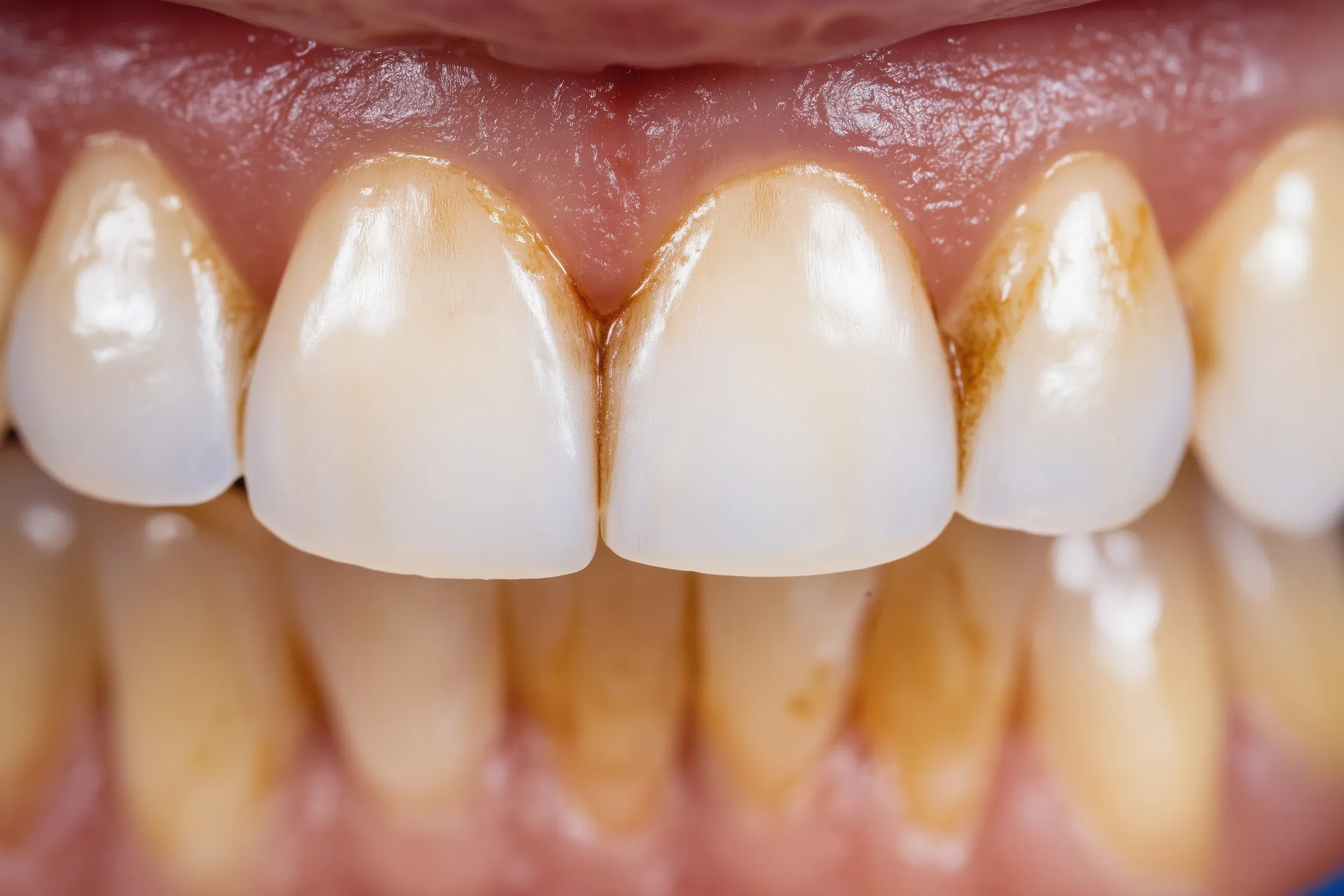
Enamel, the outermost layer of every tooth, is a highly mineralized crystalline structure that gives teeth their natural translucency. Despite being the hardest tissue in the human body, enamel is microscopically porous, and chromogenic molecules from dark beverages and foods can penetrate those pores and bond to the underlying hydroxyapatite crystals. Over time, these chromogen deposits accumulate faster than routine brushing can remove them, producing the progressively darker surface tone that characterizes extrinsic staining. According to the American Dental Association, professional cleaning and whitening are the most evidence-supported methods for safely removing these surface deposits.
Beneath the enamel lies dentin, a softer, naturally yellowish tissue that becomes increasingly visible as enamel thins with age. Starting as early as the mid-30s, enamel erodes through acid exposure and normal wear, causing the deeper dentin color to show through and shift the overall tooth tone toward yellow or gray. Simultaneously, the inner pulp chamber shrinks and darkens as it deposits secondary dentin across its walls, reducing the amount of light that passes through the tooth and making it appear dull or gray from the outside.
Intrinsic staining adds another layer of complexity. Tetracycline antibiotics taken during tooth development (roughly ages 1 through 8) incorporate directly into the dentin matrix, producing gray, brown, or banded discoloration that bleaching agents cannot fully penetrate. Fluorosis, caused by excessive fluoride intake during enamel formation, creates white spots or brown mottling within the enamel structure itself. Internal trauma triggers bleeding inside the pulp chamber; the iron sulfide compounds produced by degrading hemoglobin permanently stain the surrounding dentin from the inside out, often producing a single dark tooth that is visibly different from its neighbors.